
Modern Medical Education at JMCH: Advanced Radiotherapy, Cath Lab and Virtual Anatomy Technology
Medical education is evolving alongside advances in diagnosis, treatment and medical technology. For aspiring doctors, understanding how these technologies work is an important part of connecting classroom learning with the clinical applications of modern medicine.
At JIET Medical College and Hospital (JMCH), Jodhpur, students have an academic environment supported by a teaching hospital, specialised medical facilities and practical learning resources. The hospital's listed infrastructure includes TrueBeam and Halcyon radiotherapy systems, a Philips Azurion 7 Cath Lab and a range of academic facilities. JMCH also plans to introduce a virtual anatomy dissection table for student use from 1 December 2026.
These technologies represent three distinct aspects of contemporary medical practice: cancer treatment, interventional cardiology and digital anatomy education. Understanding their applications can help students appreciate how medical science is translated into clinical practice.
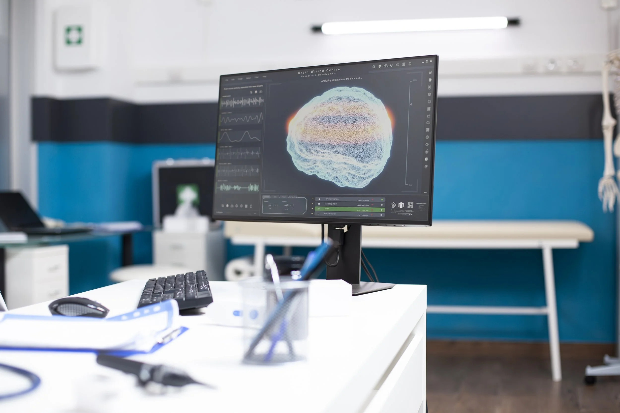
JIET Medical College and Hospital, Jodhpur, combines medical education with specialised hospital infrastructure. Its listed facilities include TrueBeam and Halcyon radiotherapy systems and a Philips Azurion 7 Cath Lab. A virtual anatomy dissection table is planned for student use from December 2026. These technologies illustrate the connection between medical science, clinical practice and contemporary healthcare education.
1. Advanced Radiotherapy: Understanding the Science Behind Cancer Treatment
Radiotherapy is an important treatment option for several types of cancer. It uses ionising radiation to damage cancer cells while treatment is planned to minimise exposure to surrounding healthy tissues.
Modern radiation oncology combines clinical assessment, diagnostic imaging, treatment planning and specialised equipment. Learning about these elements helps medical students understand the scientific principles behind cancer treatment.
JMCH lists two linear accelerator systems in its hospital infrastructure: TrueBeam and Halcyon.
TrueBeam Radiotherapy System
TrueBeam is a linear accelerator platform used to deliver radiation therapy. Its capabilities support advanced radiotherapy techniques that require detailed treatment planning and precise radiation delivery.
For medical students, understanding TrueBeam provides an opportunity to explore the relationship between medical physics, diagnostic imaging and radiation oncology.
Key learning areas include:
Understanding the role of imaging in identifying and assessing tumours.
Learning why radiotherapy treatment plans are individualised.
Understanding the importance of patient positioning and image guidance.
Exploring how radiation oncologists and medical physicists contribute to treatment planning.
These concepts connect subjects such as anatomy, pathology and oncology with the principles used in modern cancer treatment.
Halcyon Radiotherapy System
Halcyon is another linear accelerator platform listed at JMCH. It is designed to support image-guided radiotherapy workflows.
Learning about these systems helps students understand that radiotherapy involves more than delivering radiation. Treatment requires careful planning, verification, appropriate positioning and coordination among trained healthcare professionals.
The educational value lies in understanding the principles behind the technology and the responsibilities of the clinical team.
Read more about radiotherapy services at JMCH.
2. Cath Lab Technology: Exploring Interventional Cardiology
Cardiovascular medicine involves understanding the structure and function of the heart, identifying cardiac conditions and evaluating appropriate treatment options. Some conditions require specialised procedures guided by medical imaging.
A cardiac catheterisation laboratory, commonly known as a Cath Lab, is equipped with imaging technology that helps doctors visualise cardiovascular structures and guide selected interventions.
JMCH lists the Philips Azurion 7 Cath Lab among its specialised medical facilities.
What Is a Cath Lab Used For?
A Cath Lab supports procedures such as coronary angiography and angioplasty.
Coronary angiography helps doctors visualise the coronary arteries and assess possible narrowing or blockages.
Angioplasty may be performed in selected cases to improve blood flow through a narrowed coronary artery. Depending on the clinical situation, the procedure may also involve placing a stent.
The Philips Azurion 7 platform provides an example of how specialised imaging technology is used in interventional cardiology.
What Can Medical Students Learn from Cath Lab Technology?
Understanding Cath Lab procedures can help students connect cardiovascular anatomy and physiology with the investigation and management of heart disease.
Relevant learning areas include:
The structure and function of the coronary arteries.
How coronary artery disease can affect blood flow to the heart.
The role of angiography in cardiovascular assessment.
How imaging supports selected interventional procedures.
The importance of teamwork and patient monitoring during cardiac care.
Any direct observation of clinical procedures depends on the student's stage of training, the prescribed curriculum, faculty supervision and applicable patient-consent protocols.
Explore the JMCH Cath Lab facilities.
3. Virtual Anatomy Technology: A New Approach to Learning Human Anatomy
Anatomy is one of the foundational subjects in medical education. Students need to understand the structure of the human body and the relationships between organs, tissues, nerves, blood vessels and other anatomical components.
Traditional learning methods, including textbooks, anatomical models and dissection, remain important. Digital technologies can complement these approaches by allowing students to explore anatomical structures in three dimensions.
JMCH's official website lists a virtual anatomy dissection table planned for student use from 1 December 2026. This is a planned facility, and its operational availability should be confirmed through official updates before publication.
How Can Virtual Anatomy Support Medical Education?
A virtual anatomy dissection table can provide a digital environment for exploring the human body layer by layer.
Potential educational applications include:
Visualising anatomical structures in three dimensions.
Understanding the spatial relationships between organs and tissues.
Exploring complex anatomical regions from different perspectives.
Revisiting difficult concepts during revision.
Connecting anatomy with the principles of medical imaging.
For example, students studying the cardiovascular system can use three-dimensional visualisation to understand the position of the heart, its chambers and major blood vessels.
Similarly, visualising muscles, bones, joints and nerves can help students develop a clearer understanding of the musculoskeletal system.
Virtual anatomy is best viewed as a complementary learning resource. Its educational value depends on how it is integrated into teaching and supported by faculty guidance and conventional practical learning.
4. Connecting Medical Theory with Technology-Based Learning
Radiotherapy, interventional cardiology and virtual anatomy serve different purposes, but each demonstrates how scientific knowledge is applied in contemporary medicine.
Consider how these technologies relate to the MBBS curriculum:
Anatomy and Physiology: Understanding the structure and function of the human body provides the foundation for studying cardiovascular procedures and other clinical interventions.
Pathology: Learning how diseases develop helps students understand why particular investigations or treatment approaches may be considered.
Radiology and Medical Imaging: Understanding imaging principles helps students appreciate how doctors investigate conditions and guide selected procedures.
Oncology: Studying cancer biology and treatment methods provides context for understanding the role of radiotherapy in cancer care.
Clinical Reasoning: Connecting scientific knowledge with clinical examples helps students understand the reasoning behind investigations and treatment decisions.
The purpose of technology-supported education is not simply to familiarise students with equipment. It is to help them understand the scientific principles, clinical applications and limitations associated with its use.
5. Academic Facilities That Support Practical Learning at JMCH
Specialised medical equipment is only one part of medical education. Students also need appropriate academic resources, practical training environments and opportunities to develop their knowledge progressively.
JMCH's official website lists several facilities supporting medical education, including:
Lecture Theatres
Lecture theatres equipped with audio-visual facilities provide an environment for classroom teaching, presentations and academic discussions.
Skills Laboratories
Skills laboratories can support the practice of selected medical procedures using appropriate models and simulation equipment. These controlled environments help students become familiar with procedures before undertaking relevant clinical tasks under supervision.
Digital Central Library
A medical library supports textbook study, examination preparation and access to academic resources. JMCH's Central Library also lists medical journals, multimedia resources and online databases.
Departmental Learning Resources
Basic medical science departments, laboratories and museums support the study of subjects such as Anatomy, Physiology, Biochemistry, Pathology and Microbiology.
Together, these resources can help students build a foundation in medical science while preparing for later stages of clinical education.
Explore the JMCH non-clinical departments and the Central Library for further information.
6. Why Supervised Clinical Learning Matters
Advanced medical technology can support education, but the quality of learning also depends on teaching methods, faculty guidance and opportunities appropriate to the student's stage of training.
Medical students need to understand how clinical teams assess patients, interpret investigations and consider treatment options. They must also learn the importance of patient confidentiality, informed consent, ethical conduct and safety.
A structured learning environment can help students develop:
Scientific understanding: Connecting foundational medical subjects with clinical applications.
Clinical awareness: Understanding the roles of different medical specialties.
Technology literacy: Learning what medical equipment does and where its limitations lie.
Multidisciplinary thinking: Recognising how different healthcare professionals contribute to patient care.
Professional responsibility: Understanding that clinical decisions require appropriate training, supervision and judgement.
At JMCH, specialised hospital facilities provide a context for understanding these aspects of contemporary medicine. The extent of student exposure depends on the curriculum, clinical rotations and supervision arrangements.
7. The Future of Medical Education at JMCH
Medical technology continues to influence how healthcare professionals investigate diseases, plan treatments and learn new procedures.
Radiotherapy systems demonstrate the relationship between medical physics, imaging and cancer treatment. Cath Lab technology illustrates how imaging can guide selected cardiovascular interventions. Virtual anatomy offers another approach to studying the human body through three-dimensional visualisation.
JMCH's planned virtual anatomy dissection table represents a proposed addition to its academic learning resources, with student use scheduled to begin on 1 December 2026, according to the institution's website.
As the facility moves towards operation, students and prospective applicants should refer to official updates for confirmation of availability.
Ultimately, modern medical education depends on the effective integration of technology, foundational science, practical learning and responsible clinical practice.
Frequently Asked Questions
1. Which radiotherapy systems are listed at JMCH?
JMCH lists TrueBeam and Halcyon linear accelerator systems among its radiotherapy facilities.
2. What is the Philips Azurion 7 Cath Lab used for?
A Cath Lab supports image-guided cardiovascular procedures, including coronary angiography and angioplasty when clinically indicated. JMCH lists the Philips Azurion 7 system among its specialised facilities.
3. What is a virtual anatomy dissection table?
It is a digital learning platform that allows students to explore anatomical structures in three dimensions. JMCH lists a virtual anatomy dissection table planned for student use from 1 December 2026.
4. Can virtual anatomy replace traditional dissection?
Virtual anatomy can complement conventional teaching, anatomical models and dissection. Its role depends on the curriculum and the teaching methods adopted by the institution.
5. How can a Cath Lab help medical students understand cardiology?
It provides context for studying cardiovascular anatomy, coronary artery disease, diagnostic imaging and selected interventional procedures. Direct clinical observation depends on training requirements, supervision and applicable hospital protocols.
6. How does radiotherapy relate to medical education?
Radiotherapy provides an opportunity to understand the principles of radiation oncology, treatment planning, imaging and multidisciplinary cancer care.
7. Where is JIET Medical College and Hospital located?
JIET Medical College and Hospital is located at JIET Universe, NH-62, Jodhpur Pali Highway, Village Mogra, Jodhpur, Rajasthan.
Conclusion
Modern medical education brings together foundational science, practical learning and the technologies used in contemporary healthcare.
At JIET Medical College and Hospital, the listed TrueBeam and Halcyon radiotherapy systems and Philips Azurion 7 Cath Lab illustrate the role of specialised infrastructure in cancer care and cardiovascular medicine. The planned virtual anatomy dissection table represents another approach to supporting anatomy education through digital visualisation.
The value of these technologies lies in how they are integrated into structured teaching, supervised learning and the development of sound clinical judgement.
For students exploring medical education in Jodhpur, understanding the relationship between academic facilities, hospital services and specialised medical technology can be an important part of evaluating their educational options.
Visit the official JMCH website to learn more about the institution, its academic facilities and its hospital services.


